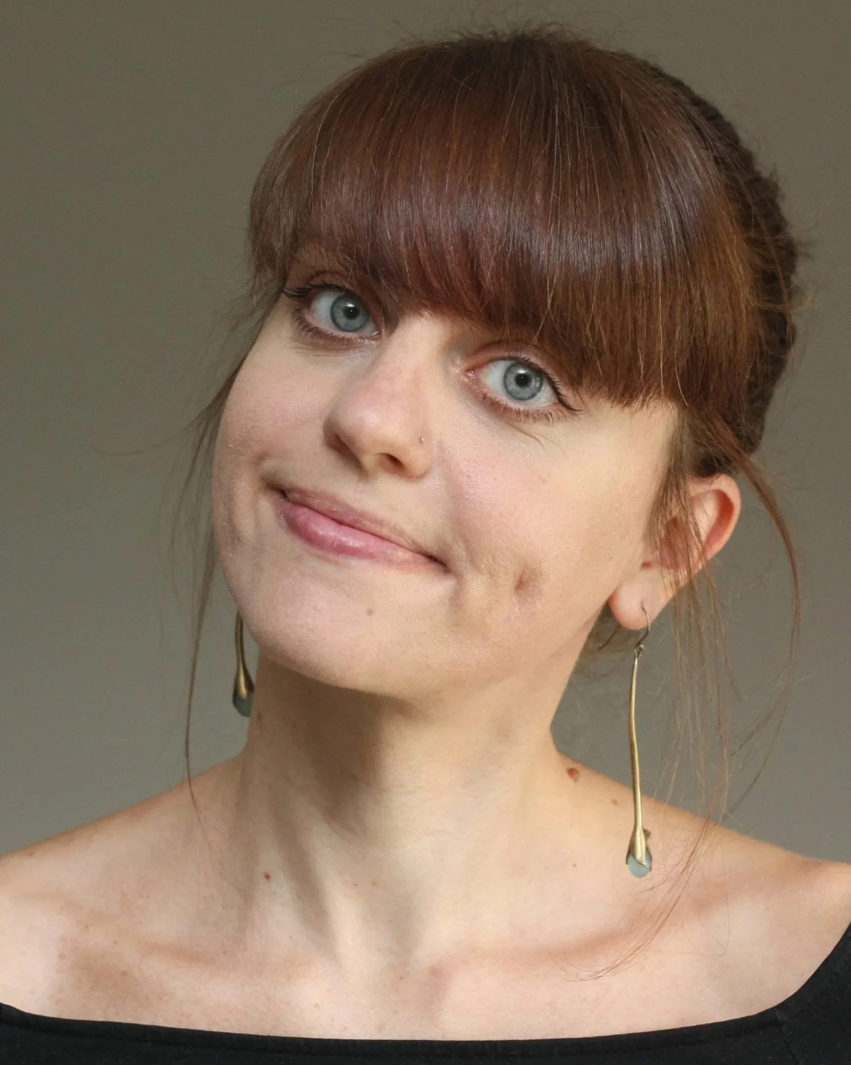
Dr. Carol Mason
Professor, Columbia University
Postdoctoral Fellow, University of Bristol and University of Wisconsin-Madison
PhD, University of California Berkeley
From collecting seashells on the Jersey Shore to fishing with her father, the natural world inspired curiosity and joy in Dr. Carol Mason from an early age. That initial curiosity led her from plant and marine biology, to zoology, to physiology and anatomy and, ultimately, to the burgeoning field of neuroscience. Over the years, Carol has uncovered essential cellular and molecular principles that guide the proper wiring of the visual system and cerebellum through development. As a Professor at the Zuckerman Institute at Columbia University and a member of the National Academies of Sciences and Medicine, her vast scientific contributions to the field are in addition to her work as an influential leader and a mentor to many.
Given her early fascination with the natural world, it is unsurprising that Carol was initially interested in biology as an undergraduate at Chatham College in Pittsburgh, a women’s college at the time. Although she didn’t much care for her first research experience in genetics, she was encouraged by a few of her favorite professors to apply to NSF-sponsored summer undergraduate research programs. She was accepted to one of these programs that allowed her to spend two summers working at the American Museum of Natural History in New York City with Dr. Dorothy Bliss to study the biology of land crabs. Carol took different scientific and life inspiration from Dorothy and from a postdoc, Dr. Linda Mantel, with whom she worked closely, and the experience convinced her to pursue research as a possible career.
As she applied to graduate schools, the cultural momentum of the late 1960’s drew Carol to Berkeley, California to continue working on invertebrate physiology. After continuing her work on land crabs for her master’s degree in zoology, she felt increasingly drawn instead to microscopy. She loved working with microscopes and relished the ability to literally see what was going on ‘under the hood’. Thus, she switched PhD advisors to study the neuroendocrine system of insects with Drs. Howard Bern and Hugh Fraser Rowell. Specifically, she used novel dyes, Procion Yellow and Cobalt Chloride, to fluorescently label and trace individual cells. These particular dyes were advantageous for rendering axonal projections in exquisite detail, allowing Carol to probe the projection pathways and targets of hormone-releasing neurons in the insect equivalent of the hypothalamus. Although her findings of previously unseen connections in this system initially ruffled some feathers, her images’ striking clarity and detail spoke for themselves and ultimately convinced prominent figures in her field.
At various points during graduate school, Carol seriously considered leaving for alternate paths that ranged from pursuing an MD to accepting an offer to work as a baker at the famed Berkeley restaurant Chez Panisse. But in the end, Carol completed her PhD and started looking for a postdoc. Interested in further exploring neuronal connectivity, she began her first postdoc with Dr. Dennis Lincoln at the University of Bristol to continue studying connectivity in the neuroendocrine system, but this time in vertebrates. Although the same tracing techniques she had used in her PhD didn’t work as well for labeling hypothalamic neurons in rats as they had in invertebrates, she happened to find that hypothalamic projections that originated in the eye were an exception. This was her first, but certainly not her last, foray into the visual system.
As much as she loved living in Europe, it was 1974, and ‘Neuroscience’ as a field was just coming into its own back in the States with the creation of the Society for Neuroscience (SfN). Drawn towards this exciting scientific frontier, Carol turned down an offer from a famous lab at Cambridge to move to Madison, Wisconsin and work with Dr. Ray Guillery. Together with another postdoc Dr. John Robson, she studied connectivity in the cat visual system using a new tool known as horseradish peroxidase (HRP), which was useful both for labeling neurons and for detecting bona fide synaptic partners through correlated light and electron microscopy. Together, they characterized the diverse morphology of projections from the retina to the visual thalamus and their form related to their function. They also discovered that these ‘retinogeniculate’ axons will ‘sprout’ new endings that defy eye-specific segregation in the thalamus if the animal loses one eye during development. From this work, and following Ray’s gentle advice and encouragement to ‘carve her own territory’ distinct from her co-postdoc, Carol began characterizing how these retinogeniculate connections form and mature during development. This established a clear scientific niche for herself and laid the groundwork for the rest of her career.
Carol began her independent research program at New York University School of Medicine in 1980 before moving to Columbia University in 1987. Initially, she focused on the development of circuit connectivity in the cerebellum before a collaboration led her back to the visual system. Her first graduate student Dr. Paula Bovolenta’s careful tracing of retinal ganglion cell projections with HRP injected to the eyes of mouse embryos revealed a remarkable diversity in the morphology of retinal neurons’ growth cones (endings of still-growing axons) on their way into the brain. Carol’s lab also described for the first time how some retinogeniculate axons cross the midline of the brain through the optic chiasm to connect contralaterally, whereas a smaller subgroup seemed to be deterred from the midline and thus are confined to the same hemisphere. In collaboration with Pierre Godement, they subsequently used video time-lapse microscopy to confirm that these axons are actively repelled from the midline. Carol and her lab then went on to explore the various molecular cues at the optic chiasm that direct whether axons are barred or permitted to cross on their way into the brain. Today, now at Columbia, Carol continues to study connectivity from the eye to the brain via the optic chiasm, with a particular focus on albinism. Although this condition is defined by lack of pigment in the hair, eyes and skin, there are also alterations to the visual system - specifically in the proportion of cells in the eye that cross or do not cross the midline, with consequences for depth perception. Her lab seeks to understand the complicated molecular landscape that regulates these varied projection patterns, how they are impacted in albinism, and how they might be targetable for intervention to improve disordered vision.
Even as Carol was pioneering early connectomics and making foundational discoveries in the visual system, she was also navigating personal challenges. She met her husband while she was at NYU and he was a professor at Yale, and their two-body problem only became more complicated from there. First, she found out that she was pregnant with her first child just as her husband was offered a position across the country at Stanford. Then years later, after he had a faculty position at Yale, it happened again – he was offered a position at Princeton, and she was pregnant once again in her mid-40s. Balancing parenthood with work through constant inter-state commutes was a regular challenge, although silver linings emerged as well. For instance, during the extensive time she spent visiting her husband in the Bay Area while pregnant with her first child, she made close friends and colleagues and gained new scientific inspiration. Meanwhile, Carol also had to navigate being a woman in science at a time when there still were frustratingly few. She too often observed and/or experienced different treatment, such as when she and another female colleague were excluded from discussions with institutional leaders about what it would take to keep them from moving institutions – discussions that only involved her male colleagues until she pushed to be included.
Even though Carol describes herself as somewhat ‘conflict-averse’, she is also someone who argues for change when she sees something she doesn’t like. In doing so, she has emerged as an important leader and advocate in the field. As a graduate student at Berkeley, she lobbied for (and succeeded in) getting students in the room for faculty meetings, and more recently has facilitated bringing junior faculty into executive meetings at her own institution. She is currently the Zuckerman Institute’s Chair of Interschool Planning and has previously served as the President of the Society for Neuroscience and as the Director of Columbia’s neurobiology graduate program, among other roles. She reflects, “I’ve always tried to change things to be more inclusive and better for trainees or the field, but I haven’t always known the right way to do that.” Although she notes that she may not always be as vocal as some of her colleagues and never had any formalized leadership training, she has instigated change, successfully, in her own way. In so doing, Carol has amassed a remarkable legacy – not just in discovering fundamental principles of the visual system that fill our textbooks, but also through her personal approach to leadership, mentorship, and advocacy.
Find out more about Carol and her lab’s research here.
Listen to Megan’s full interview with Carol on August 6, 2025 below!